Breast cancer
Symptoms and causes
Symptoms and causes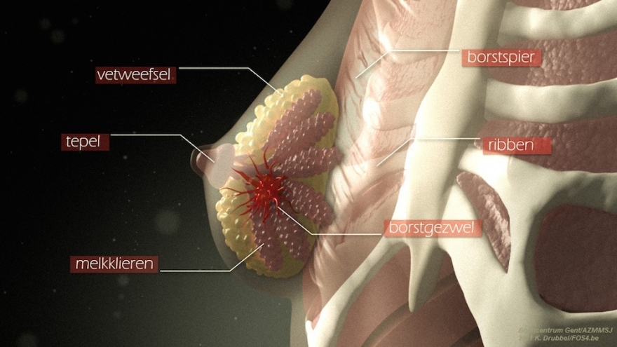
What is it?
Breast cancer is the malignant degeneration in the breast gland tissue. Internally, the breast consists mainly of glandular, binding and fatty tissue.
The glandular tissue contains about twenty mammary glands. Each of these glands drain via a duct under the nipple. They are separated by connective tissue partitions. In most cases, degeneration occurs in the milk ducts, less frequently in the glandular tissue itself. Tumours on the nipple, in the skin, in the muscle cells under the nipple, in connective tissue and in blood and lymph vessels are more rare.
Diagnosis and treatment
Diagnosis and treatmentDiagnosis
In order to be able to notice a change in time yourself, it is a good idea to check your breasts regularly at any age.
There are several starting points.
Usually, a suspicious lesion is found during routine screening. Small calcifications (microcalcifications) are then often seen. To find out more, it is best to perform a puncture biopsy. This means that the physician will remove tissue cells using a thin needle. After microscopic examination of that tissue, it becomes clear whether or not the lesion is malignant.
The biopsy can be done by ultrasound (when the lesion or calcifications can be observed under ultrasound) or by a stereotactic biopsy.
When a lesion is suspicious, clinically or radiologically, it is best to perform a puncture biopsy. This means that the physician will remove tissue cells using a thin needle. After microscopic examination of that tissue, it becomes clear whether or not the lesion is malignant.
Besides the clinical examination and the mammography, the physician also examines the structure and cells of the nipple fluid and a galactography is performed. That is an examination of the nipple gland and milk ducts. Afterwards, the observed lesion is removed and examined microscopically. If it is malicious, a balance is drawn up for further discussion.
In case of a skin lesion of the nipple, a mammogram and best preferably als an NMR examination will be performed. Additionally, a piece of tissue from the lesion or nipple will be removed under local anaesthetic (biopsy).
When one or more glands in the armpit are enlarged, even if a further examination and palpation of the breast is normal, it is best to perform a puncture of this gland with a fine needle. This will allow for the examination of tissue cells.
In case of redness and/or fluid accumulation (oedema), there is a distinction between an infection and an underlying (extensive) malignant process.
NMR examination and tissue examination can help to make the correct diagnosis through a biopsy.
Further testing
Further testingMedical imaging
The goal of imaging is to detect as many breast tumours as possible at an early stage. In doing so, we aim to minimise side effects as much as possible onthe patient' s physical and mental well-being . The three main imaging techniques are:
These three techniques work in completely different ways. That means the physician collects tissue cells through a thin needle. After microscopic examination of that tissue, it is becomes clear whether or not the lesion in the mammary gland and breast cancer is malignant.
Tissue examination
Tissue examination is essential to diagnose cancer. The pathologist is the specialist who determines whether there is cancer or not, by means of tissue analysis in the lab. For this, she needs a piece of representative tissue (biopsy). This can be obtained in several ways.
Staging examinations
Once the diagnosis of breast cancer has been confirmed, the attending physician will request additional 'staging examinations'. This means that the physician will determine what stage the cancer is in so that the spread of cancer cells (metastases) can potentially be ruled out.
Read more here about the different staging examinations.
Treatment
TreatmentNot every patient receives the same medical treatment. It all depends on your diagnosis, the degree of malignancy and stage of the cancer, the scientific evidence of treatment effectiveness for this type of cancer and your general health condition. Your physician will map out everything you need to choose the most appropriate treatment.
Surgery
Most patients need surgery to remove their tumour. Two surgeries are possible for breast cancer treatment: a breast-conserving operation or a complete removal of the breast. Sometimes, the lymph nodes in the armpit are also removed during the operation. This is because the cancer often spreads through the lymph nodes. It is therefore important to check whether the lymph nodes also contain cancer cells.
Here you can consult information on all possible breast cancer operations and the possibility of breast reconstruction.
Complementary therapies
Treatment centres and specialisations
Treatment centres and specialisations


Latest publication date: 14/05/2024
Supervising author: Dr Elzo Kraemer Ximena, Dr De Craene Annick