Supine bicycle echocardiography
What is it?
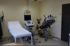
What is it?Sometimes, an echocardiography at rest provides insufficient information about the heart because specific abnormalities only manifest during cardiac exercise. During a supine bicycle echocardiography, an echocardiography is performed during physical exertion. The physical exertion is achieved by cycling in a supine position on a special recumbent bicycle.
What is the process?
What is the process?Preparation
No specific preparations are required. Make sure you wear comfortable clothes and shoes that allow for easy movement. After having removed your upper clothing, you will take place on the recumbent bicycle in a semi-reclined position. ECG electrodes are attached to your chest to monitor your heart rhythm. A blood pressure monitor is put around your upper arm.
You are secured onto the bicycle with a belt around your waist. To ensure proper imaging of the heart, you will be turned slightly onto your left side.
Test
You will be asked to cycle. The bicycle resistance is gradually raised. At every stage, the cardiologist will take images of the heart using an ultrasound device. Please tell the cardiologist or nurse if you are unable to cycle any further.
If maximum exertion has been reached, you will be asked to continue cycling for a few more minutes without resistance. During this recovery phase, pulse rate and blood pressure are allowed to gradually drop to their initial values.
Aftercare
You may feel slightly tired after the test. The heart as well as the rest of the body has engaged in exercise.
What are the risks?
What are the risks?You may feel short of breath after the exertion. This is normal and generally resolves by itself. Let the physician know if you feel pain or pressure in your chest.
You will be monitored continuously during the procedure by means of the electrocardiogram and the blood pressure monitor. These measurements allow for the timely detection of any heart rhythm disturbances. Equipment and medication to treat any potential heart rhythm disturbances are always available.
Results
ResultsAt the end of the consultation, the cardiologist will discuss the findings of the ultrasound with you as well as any resulting changes in your treatment.
Centres and specialist areas
Centres and specialist areas
Latest publication date: 16/05/2024
Supervising author: Dr Provenier Frank