Echocardiogram through the chest
What is it?
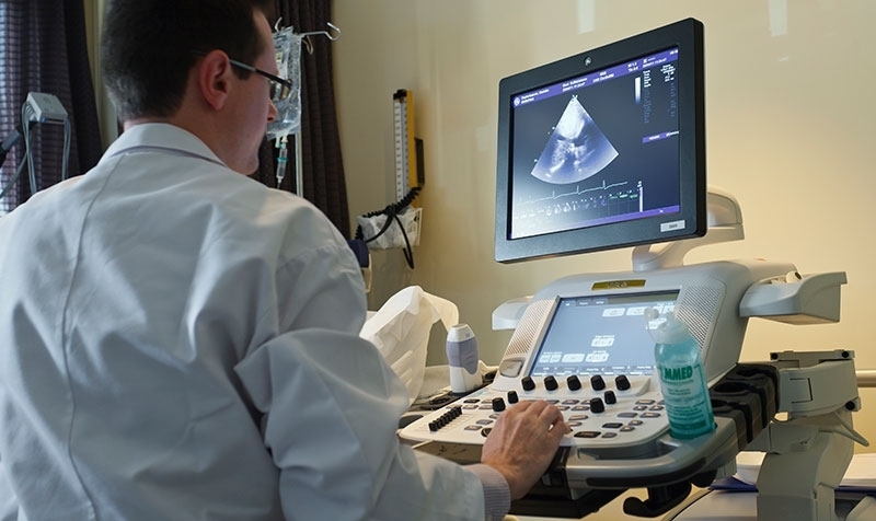
What is it?An echocardiogram is a test that uses sound waves to examine the structures and functioning of the heart. This test provides information on the dimensions about the different chambers of the heart, wall and valve movement and about any abnormalities.
The sound waves are emitted and received by a transducer or probe. Through the reflection of the sound waves, moving images of the heart and the heart valves are formed. Depending on the materials used, this can be a 2D or 3D image.
A Doppler test is performed in conjunction with the echocardiography. This is used to test the velocity of the blood flow through the different valves.
What is the process?
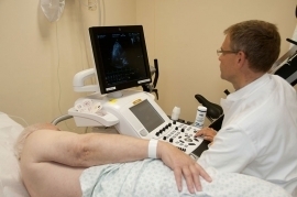
What is the process?Preparation
No specific preparations are required. After having removed your top and underwear, you will take place on the treatment table. Three electrodes on your chest are used to monitor your heart rhythm. To ensure proper imaging of the heart, you will be asked to lie on your left side. The room will be darkened to allow the cardiologist the best possible view of the echocardiogram.
Procedure
To ensure good conduction of the sound waves, a cold gel is applied to the chest. The probe is placed on various places on the chest wall to be able to view the heart from all sides. During the examination, you may be asked to hold your breath for a few seconds a few times. The examination is pain free and the only thing you will notice is the vibrations produced by the probe.
It will take up to 30 minutes. An ultrasound of the heart through the chest is not associated with any risk and does not require any specific aftercare.
Results
ResultsAt the end of the consultation, the cardiologist will discuss the ultrasound findings with you as well as any resulting changes in your treatment.
Centres and specialist areas
Centres and specialist areas

Latest publication date: 16/05/2024
Supervising author: Dr Provenier Frank