Lymph oedema
Symptoms and causes
Symptoms and causesWhat is it?
The lymphatic system is a transport system that works closely with blood circulation. It is made up of lymphatic vessels and is comparable to vascular structures. The lymphatic system ensure that the lymph fluid is disposed of.
Lymph nodes are where the large lymph vessels intersect. Because the lymphatic system and the blood vessels are in contact with each other, substances that are present in the lymphatic fluid can be excreted.
The lymphatic system has the following functions:
- dispose of excess fluid that contains waste materials from the tissues
- fight infections
- produce lymph from tissue fluid
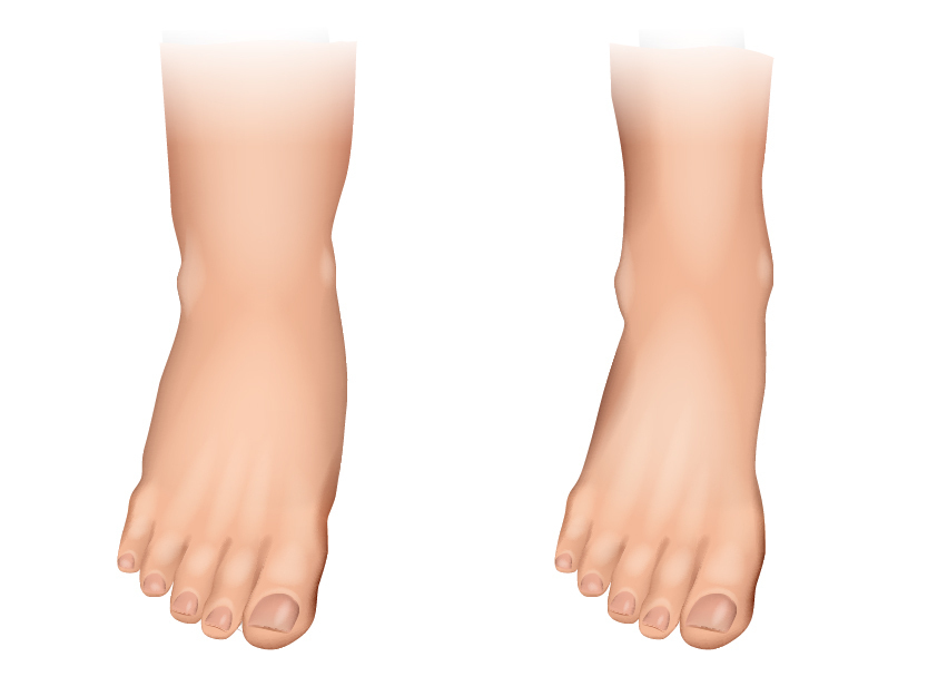
Lymphoedema is an lack of fluid discharge via the lymphatic pathways with an abnormal accumulation of protein and fluid in body tissue as a result. Over time, the lymphatic vessels that still work wear out and they stop functioning. As a result, the valves in the lymphatic vessels are affected and leak lymphatic fluid out of the lymphatic vessels due to elevated pressure. This, in turn, causes skin swelling.
Lymph oedema usually happens in the legs and/or arms, but it can also happen in the head, neck, chest, abdomen and pubic areas.
Symptoms
General characteristics
General characteristics of lymph oedema include:
- swelling
- fatigue and a heavy feeling in the affected extremity
- tight, painful feeling of the skin
- lightly-discoloured, smooth skin
- limited mobility
- later stage: skin abnormalities and tissue build-up in the lymphatic vessels
- infections
Characteristics per stage
Lymph oedema presents in various stages. See below for an overview of the symptoms of each stage:
The lymphatic vessel system is already damaged in this stage, but there are not any symptoms yet because the outflow of lymph can still be compensated for by the undamaged portion of the lymphatic vessel system.
The lymphatic vessel system is damaged and its function is damaged. The affected body parts have increasing swelling over the course of the day. The tissue swelling is still mild, such that you can still easily make an 'indent': pitting oedema. The swelling is able to disappear if you elevate your leg.
With the accumulation of protein-rich lymph, the subcutaneous tissues become affected: proliferation (fibrosis) and hardening (sclerosis) of the tissue takes place. Elevating the area does not reduce the swelling. The oedema becomes non-pitting: indents can no longer be made in the skin. There are also skin changes.
More subcutaneous connective tissue forms making the skin even tighter. The swelling takes on enormous proportions. There can be thickening as well as wart-like proliferations. The skin changes are also very pronounced. The skin is very sensitive and, because it is hard for wounds to heal, it is also very susceptible to infections such as erysipelas.
Origin
Primary lymphoedema
Cogenital lymph oedema, or primary lymph oedema, originate from either an insufficient construction lymphatic vessels or from ill-functioning lymphatic vessels. It may be evident at birth or only years later, when the intact portion of the lymph system becomes over-stressed. Primary lymph oedema often ends up affecting both sides. Most types of lymph oedema are not hereditary, although there can be a family tendency.
Secondary lymph oedema
Secondary, or acquired, lymph oedema is much more common. It can originate from a trauma, surgical procedure, radiation or infection. A very common form of lymph oedema in the arm arises after an operation for breast cancer, where the lymph nodes have been removed from the underarm. Lymph oedema in the leg often occurs after a surgical intervention for varicose veins or after treatment for gynaecological tumours.
Risk factors
It is often a progressive disease that gets more and more common the older someone becomes, and when certain risk factors play a role:
- smoking
- alcohol
- being overweight
- insufficient activity or movement
- a (prior) skin infection such as erysipelas
- insect bites
- contusion with haematoma
- obstructed discharge from the lymph, such as tight clothes
- thrombosis in an arm or leg
When to see a physician?
If you suspect lymph oedema, it is important to consult a physician. As described above, treating lymph oedema in an early stage is important because the results are much better than treating it at a later stage: 'prevention is better than healing (too late)'. It is important that the advanced skin and subcutaneous changes be prevented because they are irreversible.
Diagnosis
The diagnosis of lymph oedema is made based on the basis of:
1. A physician's clinical examination
2. Volume measurement of the extremity
3. Imaging
- Doppler duplex testing
- MRI scan
- CT scan
- Spectroscopy
With a spectroscopy, electrodes send non-liquid current through the skin to detect the presence of tissue fluid outside of the cells.
4. Lymph fluoroscopy
After subcutaneous injection of a fluorescent substance, a camera visualises the accumulation of the superficial lymph system. This allows us to optimise treatment.
5. Lymph scintigraphy
A lymph scintigraphy visualises the lymphatic vessels in the arms and legs. We can see if there is a question of lymph oedema, and, if so, how extensive it is.
The most common tests are a Doppler duplex test and a lymph scintigraphy.
Treatment
Early treatment of lymph oedema is important, especially to prevent subsequent complications. By treating as early as possible and reducing the oedema, the skin and subcutaneous tissue changes will be counteracted and infections can be prevented.
Treatment of lymph oedema does require a lot of discipline and perseverance from the patient.
Treatment is different for pitting and non-pitting oedema. With pitting oedema, the oedema can be reduced in scope. There is first a shifting phase and then a maintenance phase. With non-pitting oedema, shifting is no longer possible: we proceed right into the maintenance phase.
Without treatment, the oedema usually increases due to scarring of the skin and subcutaneous tissue. It becomes susceptible to infection. These infections then cause further damage to the skin, subcutaneous structures, and lymphatic vessels.
Conservative treatment
Surgery
We do not immediately operate for lymph oedema. Surgery is considered only if the combination of previous therapies have given an insufficient result. Possible surgical techniques are:
- liposuction: suctioning of the subcutaneous fat and lymph fluid
- lympovenous shunt: place a connection between the lymph vessels and vascular system
- lymph node transplant
- debulking and reduction surgery
- reconstructive methods
Treatment centres and specialisations
Treatment centres and specialisations
Latest publication date: 15/05/2024
Supervising author: Dr Willaert Willem