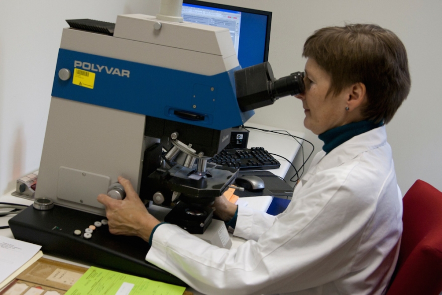
Tissue examination
Ultrasound-guided biopsy
Ultrasound-guided biopsyA core biopsy is performed with ultrasound guidance. This biopsy is performed in order to remove tissue cylinders from a wound. The tissue may then be analysed.
An ultrasound is first performed to visualise the biopsy site in order to find the easiest way to enter with the needle. The skin is disinfected and local anaesthesia is administered. A small incision allows the biopsy needle to be guided right before the site to be biopsied. Once the tissue to be analysed has been located, you will hear a click. Usually an attempt is made to take three biopsies. After the biopsy has been taken, the incision is closed with strips, which are then covered by a plaster. After two to three days, the strips can be removed.
The biopsied tissue is sent to the laboratory and the result is available within a few days.
Stereotactic biopsy
Stereotactic biopsyA stereotactic core biopsy is primarily performed with a mammography machine. The procedure can be performed sitting or lying down.
Digital X-rays are taken around the lesion in the breast. The biopsy site will be based on these images. Once the site has been determined, the radiologist will administer local anaesthesia to this area. The rest of the procedure is painless. A small incision is made to insert the biopsy needle. You will hear a click when the biopsy (e.g. the tissue to be analysed) has been taken. Two to three biopsies are usually taken. After the biopsy has been taken, the incision is closed with strips, which are then covered by a plaster. After two to three days, the strips can be removed.
The biopsied tissue is sent to the laboratory, and the result is available a few days later.
Centres and specialist areas
Centres and specialist areas


Latest publication date: 15/12/2023
Supervising author: Dr Elzo Kraemer Ximena, Dr Coppens Mieke