Kidney biopsy
What is it?
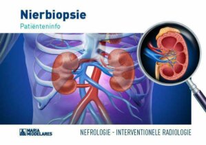
What is it?A kidney biopsy is a procedure in which a special needle (biopsy needle) is used to remove small pieces of tissue from one kidney. This is done by inserting a needle into the kidney through the skin, under a local anaesthetic. The pieces of tissue are examined under the microscope in the laboratory. Through this examination, the nature and severity of your kidney problem can be determined and the right treatment can be started.
The needle is inserted into the kidney under ultrasound guidance and, in exceptional cases, under CT guidance. These techniques facilitate the visualisation of the kidney and the needle. The procedure itself takes place at the Surgery Department and is performed by an interventional radiologist (i.e. a radiologist who is specialised in performing minimally invasive procedures) under imaging guidance.
Preparation for the procedure
Preparation for the procedureYou will be asked about your use of medication during the consultation with the nephrologist (i.e. the kidney specialist). It is important that your physician knows if you take blood thinners (e.g. Marevan®, Sintrom®, Plavix®, Asaflow®, Cardioaspirine®, Xarelto®, Eliquis®, Pradaxa®). In consultation with the nephrologist, the blood thinners will be stopped a few days before the kidney biopsy. This is done to prevent possible bleeding after the biopsy. Other required medication will also be discussed. You will be able to continue taking some medications.
Are you allergic to certain substances (medications, latex, contrast agent)? Do not forget to mention this to your nephrologist during the consultation and on your admission.
If you are pregnant, make sure to let the nephrologist know. Biopsies during pregnancy are only performed in exceptional cases.
It is important that you have blood samples collected at the GP or nephrologist surgery to check for clotting. Your nephrologist or interventional radiologist can only perform the biopsy once the results of the blood tests have been received.
Course of the procedure
Course of the procedureOn the day of the procedure, you may take a light breakfast since the biopsy is usually performed in the afternoon. You may no longer eat anything beginning from four hours before the procedure. You may drink water up to two hours before the procedure.
In the hospital, a nurse will ask you for certain details and note these down. Blood pressure, pulse rate and temperature will be measured and if required, final blood samples will be drawn. You will be given a gown by the nurse to wear in your room (you may keep your underpants on). If necessary, you will receive medication to relax.
Fifteen minutes before the procedure you will be transported, in your bed, to the radiological intervention room in the Surgery Department. You will be asked to lie on your right or left side. The procedure starts with an ultrasound examination of the kidney to determine a good puncture site on the skin. If necessary, a few pillows will be placed under your side to make it easier to insert the needle into the kidney. The skin is disinfected and a sterile sheet is placed. Then, a local anaesthetic is applied to the skin and underlying area up to the kidney, under ultrasound guidance. This is associated with a burning sensation that disappears after a few seconds.
Almost immediately after this, the needle is placed and a few (usually three) pieces of tissue
(i.e. biopsies) are taken from the underside of the kidney. This is usually pain-free; you only hear a clicking sound when the tissue sample is taken.
After the biopsy, pressure is applied to the puncture site and before you leave the operation room, a final ultrasound check is performed.
After the procedure
After the procedureAfter the procedure, and if there are no problems, you may eat and drink something after approximately an hour. You will remain in bed for a maximum of 24 hours, lying on your back. You may not get up until after the ultrasound check-up and blood draw the following morning. A sandbag is placed over the puncture site for up to six hours after the procedure. These measures are necessary to minimise the risk of bleeding at the puncture site.
The nurse will come to your room to carry out regular checks of your blood pressure and pulse rate. The dressing that has been applied to the puncture site will also be checked regularly. To prevent bleeding, you may not get up to visit the toilet. If required, ask for a bedpan or urinal. The first urine may be coloured pink or red. That is normal and is caused by the needle being inserted into the kidney.
If you feel pain or discomfort, always inform the nurse. You will stay one night in hospital for further observation and checks. The morning after the biopsy, an ultrasound will be performed at the Radiology Department to check your kidneys. Blood samples will be taken at the ward. Then, the attending nephrologist decides if you may leave the hospital.
Before your hospital discharge, a date for your next consultation with the nephrologist is agreed. During this consultation, the results of the biopsy and any further treatment is discussed.
Guidelines for at home
Guidelines for at homeStrenuous effort must be avoided in the first week after the biopsy. If, after your discharge from hospital, you have any of the following:
- fever
- pain around the biopsy site
- pinkish-red urine
- general malaise
then please contact the Nephrology Department or, if outside office hours, the A&E of the hospital.
Leaflet
LeafletMore information about kidney biopsies and possible complications can be found in the leaflet below.
Only available in Dutch:
Nierbiopsie
DownloadCentres and specialist areas
Centres and specialist areas

Latest publication date: 23/01/2024
Supervising author: Dr Schoofs Christophe, Dr De Vleeschouwer Mieke